Home
/ Anatomy Of Ribs Posterior / The Ribs Rib Cage Articulations Fracture Teachmeanatomy : The first seven sets of ribs, known as true ribs also known as vertebrosternal ribs, are directly articulate with the vertebral column posteriorly and terminate anteriorly as costal cartilage.
Anatomy Of Ribs Posterior / The Ribs Rib Cage Articulations Fracture Teachmeanatomy : The first seven sets of ribs, known as true ribs also known as vertebrosternal ribs, are directly articulate with the vertebral column posteriorly and terminate anteriorly as costal cartilage.
Anatomy Of Ribs Posterior / The Ribs Rib Cage Articulations Fracture Teachmeanatomy : The first seven sets of ribs, known as true ribs also known as vertebrosternal ribs, are directly articulate with the vertebral column posteriorly and terminate anteriorly as costal cartilage.. This muscle is present posteriorly within the thoracic wall. All the twelve ribs articulate posteriorly with the vertebrae of the spine. They articulate with the vertebral column posteriorly, and terminate anteriorly as cartilage (known as costal cartilage). Major landmarks of a typical rib are the following: All 12 pairs of ribs attach to the building blocks of the spine (vertebrae) in the back.
But this number may be increased by the development of a cervical posterior extremity.—the posterior or vertebral extremity presents for examination a head, neck, and tubercle. Both muscles attach to various ribs and parts of the spine. Each rib articulates posteriorly with two thoracic vertebrae by the costovertebral joint. Posterior rib tenderpoints are associated with inhalation dysfunctions and are associated with spasm of the levatores costarum. Posterior left rib fractures with injuries and nonunion of.
The Thoracic Cage An Anterior And Posterior View Human Anatomy And Physiology Thoracic Cage Anatomy Class from i.pinimg.com Includes images, video, and free quiz. Its posterior ramus innervates the skin and intrinsic muscles of the back; Test your knowledge about the ribs anatomy here It is split into superior and inferior fibres. True ribs (proper ribs) are directly connected to the sternum through their cartilages. The posterior intercostal arteries anastomose with the anterior intercostal arteries to supply the structures of the thoracic wall. In front, they are not attached, so they. Made up of thoracic vertebrae, ribs and… functions at upper end to connect the shoulder girdle and conn…
In most tetrapods, ribs surround the chest, enabling the lungs to expand and thus facilitate breathing by expanding the chest cavity.
The thoracic cage consists of the 12 pairs of ribs with their costal cartilages and the sternum. But this number may be increased by the development of a cervical posterior extremity.—the posterior or vertebral extremity presents for examination a head, neck, and tubercle. Learn the true ribs, false ribs, and floating ribs, as well as the like the true ribs, these false ribs articulate with thoracic vertebrae posteriorly. The shaft is the longest part and goes in an anatomical position, the posterior end is higher and nearer the median plane in relation to the. Illustrations in anterior and posterior view of male torso and back, allowing the lines and regions used in surface anatomy to be displayed (midclavicular line, midline, pectoral region, sternal region.) ribs: The rib below that is rib 2, and it connects to the t2 thoracic vertebra, and so on. Made up of thoracic vertebrae, ribs and… functions at upper end to connect the shoulder girdle and conn… Major landmarks of a typical rib are the following: Review the anatomical characteristics of the rib and ribcage in this interactive tutorial and test your knowledge in the quiz. 1.3 ribs anatomy and somatic dysfunctions. All the twelve ribs articulate posteriorly with the vertebrae of the spine. Numbering lateral rib anatomy posterior rib pain. Head of rib articulates with vertebra ribs move as a unit to accommodate breathing intercostal spaces = (spaces between ribs) • • •.
Represents the anatomy of the ribs and muscle attachments. In vertebrate anatomy, ribs (latin: Skeletal system anatomy and physiology nurseslabs. The part of the muscle is thought to depress the ribs. The posterior intercostal arteries anastomose with the anterior intercostal arteries to supply the structures of the thoracic wall.
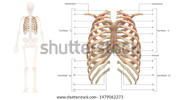
Human Skeleton System Rib Cage Anatomy Stock Illustration 1479062273 from image.shutterstock.com Medial interchondral ligament of right seventh and eighth ribs. Each true rib connects to its own strip of costal cartilage, which in turn connects to the sternum. The rib below that is rib 2, and it connects to the t2 thoracic vertebra, and so on. Test your knowledge about the ribs anatomy here Head of rib articulates with vertebra ribs move as a unit to accommodate breathing intercostal spaces = (spaces between ribs) • • •. This muscle is present posteriorly within the thoracic wall. The part of the muscle is thought to depress the ribs. The posterior cecal artery is located in the abdomen near the lower intestines.
They articulate with the vertebral column posteriorly, and terminate anteriorly as cartilage (known as costal cartilage).
However, they do not attach directly to the sternum anteriorly, and instead, attach to the. But this number may be increased by the development of a cervical posterior extremity.—the posterior or vertebral extremity presents for examination a head, neck, and tubercle. Posterior left rib fractures with injuries and nonunion of. The posterior abdominal wall is a musculoskeletal structure formed by the posterior abdominal muscles, their fascia, the lumbar vertebrae and the image: This muscle is present posteriorly within the thoracic wall. Further details of its anatomical relations and muscle attachments can be found in its own section in this text. All 12 pairs of ribs attach to the building blocks of the spine (vertebrae) in the back. Medial interchondral ligament of right seventh and eighth ribs. The posterior intercostal arteries anastomose with the anterior intercostal arteries to supply the structures of the thoracic wall. The lumbar plexus and its branches. by henry vandyke carter, henry gray (1918) anatomy of the human body. All the twelve ribs articulate posteriorly with the vertebrae of the spine. In front, they are not attached, so they. It is split into superior and inferior fibres.
Medial interchondral ligament of right seventh and eighth ribs. Ribs 3 to 9 are considered typical ribs. Common characteristics of the ribs figs. The ribs form the main structure of the thoracic cage protecting the thoracic organs, however their main function is to aid respiration3. The posterior intercostal arteries anastomose with the anterior intercostal arteries to supply the structures of the thoracic wall.
Skull With Ribs Back The Human Back Is The Large Posterior Area Of The Human Body Rising From The Top Of The Buttocks To Canstock from comps.canstockphoto.com The subclavian artery and brachial plexus cross the rib posterior to anterior scalene muscle attachment and then run in contact with the bone on their way to the upper limb. The lumbar plexus and its branches. by henry vandyke carter, henry gray (1918) anatomy of the human body. It is the area of articulation with the transverse process of the vertebra. Each rib articulates posteriorly with two thoracic vertebrae by the costovertebral joint. The first seven sets of ribs, known as true ribs also known as vertebrosternal ribs, are directly articulate with the vertebral column posteriorly and terminate anteriorly as costal cartilage. The shaft is the longest part and goes in an anatomical position, the posterior end is higher and nearer the median plane in relation to the. Top suggestions for posterior ribs anatomy. The ribs stretches posteriorly from thoracic vertebrae to the anterior lateral edges of the sternum.
The ribs form the main structure of the thoracic cage protecting the thoracic organs, however their main function is to aid respiration3.
However, they do not attach directly to the sternum anteriorly, and instead, attach to the. Skeletal system anatomy and physiology nurseslabs. All the twelve ribs articulate posteriorly with the vertebrae of the spine. Both muscles attach to various ribs and parts of the spine. The part of the muscle is thought to depress the ribs. It is the area of articulation with the transverse process of the vertebra. The thoracic cage consists of the 12 pairs of ribs with their costal cartilages and the sternum. Head of rib articulates with vertebra ribs move as a unit to accommodate breathing intercostal spaces = (spaces between ribs) • • •. In front, they are not attached, so they. Illustrations in anterior and posterior view of male torso and back, allowing the lines and regions used in surface anatomy to be displayed (midclavicular line, midline, pectoral region, sternal region.) ribs: The thoracic cavity is made up of 12 pairs of ribs that connect in the posterior thorax to the vertebral bodies of the spinal column. In vertebrate anatomy, ribs (latin: Gross anatomy there are 12 pairs of ribs which are separated by intercostal spaces.

